Main Principles of the Method and Physical Characteristics.
Ultrasound is high-frequency fluctuations that lie in the rate higher than those, perceived by a human ear (more than 20 000 Hz). Being radiated into the patient's body, ultrasonic fluctuations are reflected from the investigated tissues, blood and surfaces, such as the borders between organs, and, coming back to ultrasound scanner, are processed and measured after their preliminary delay to receive a focused image. The result data enter monitor screen that allows evaluation of internal organs condition. Even though ultrasound does not effectively penetrates through such environments as air and other gases as well as through bones, it is widely applied when investigating soft tissues. Usage of ultrasound gels and other liquids both improves probes characteristics and extends applications of ultrasound scanners for various medical examinations. The average velocity of ultrasonic waves in human soft tissues is 1.540 m/sec and is practically independent of frequency. A probe is one of the basic components of diagnostic systems that converts electric signals into ultrasonic fluctuations and products electric signals, receiving the reflected echo from internal tissues of the patient. The ideal probe must be efficient as a radiator and sensitive as a receiver, have good characteristics of the impulses radiated by it with strictly determined indeces as well as receive a wide range of frequencies reflected from the examined tissues. In electronic probes ultrasonic fluctuations are stimulated due to delivery of high-frequency pulses to piezo-crystals, that form the probe (piezo-electric effect was discovered by Pier and Mary Cury in 1880). The amount of crystal vibrations per a second determines probe frequency. With frequency increase, the wavelength of generated fluctuations decreases which causes improvement of resolution, however, the absorption of ultrasonic fluctuations by body tissues is proportional to frequency increase, that leads to the reduction of penetration depth. Thus, probes with high frequency of fluctuations provide better image resolution while investigating superficial tissues, as well as low-frequency probes allow examination of organs located deeper though image quality is lower than of high-frequency probes. This discord is the main determining factor when using probes. In everyday clinical practice various constructions of probes are used: those, that consist of discs with one element and combining some elements, located on the circumference or along the probe, producing different image formats, which are necessary or preferable while diagnosing various organs.
Traditionally and basically 5 probes types are used:
- Mechanic Sector Probes.
- Annular Probes.
- Linear Probes.
- Convex Probes.
- Probes with Phazed Scanning.
- method of ultrasonic fluctuations formation;
- method of radiation;
- created image format on the monitor screen.
 |  |  |  | |
| Phazed Probes | Linear Probes | Convex Probes | Mechanic Sector Probes | Annular Probes |
With diagnostic aims usually probes with frequencies 3.0 MHz, 3.5 MHz, 5.0 MHz. 6.5 MHz and 7.5 MHz are used. Besides, units equipped by high-frequency probes of 10-20 MHz appeared on the market of ultrasound equipment recently.
Probes Applications.
- 3.0 MHz (convex and sector) probes are used in cardiology;
- 3.5 MHz (convex and sector) probes are used in abdominal diagnostic and when examining organs of a small pelvis;
- 5.0 MHz (convex and sector) probes are used in pediatrics;
- 5.0 MHz probes with short phocus may be applied for mamma examination;
- 6.0-6.5 MHz (convex, linear, sector, annular) probes are used in the examination of cavities;
- 7.5 MHz (linear, probes with water tip) are used when examining superficial organs- thyroid gland, mammae, lymphatic system. Main parameters of image adjustment.
- Gain - "reinforcement" of the detected signal on account of variation of the relation between ranges of input and output signal. (Excessively high level of reinforcement causes indistinct image that becomes "white").
- Dynamic range - range between registered signals with maximum and minimum intensiveness (The wider it is, the better we perceive signals that have little difference in intensiveness).
- Contrast- characterises ability of the system to distinguish between echosignals with little difference in amplitude or brightness.
- Focusing - is used to improve resolution in the concrete investigated area. (Increase of the number of focus zones increases image quality but decreases cadre frequency).
- TGC - reinforcement, compensated in depth.
- Frame average - allows smoothing image due to putting of a certain number of cadres onto one another during time unit or making it hard, approaching to the real time scale.
- Direction - changes image orientation on the screen (right-to-left or top-to-bottom).
Image Artefacts.- Reverberation. Is observed in the case when an ultrasound wave occurs between two or more reflecting surfaces, partly being repeatedly reflected. At that the screen will show non-existent surfaces, which will be located behind the second reflector at the distance equal to the distance between the first and the second one. Most often it occurs when the ray passes structures that contain some liquid.
- Mirror Artefacts. Means that the image shows an object, located from one side of a strong reflector, from its other side. This phenomenon often appears near diaphragm.
- "Comet Tale". Small echopositive signals, appearing behind gas bubbles and caused by their own fluctuations.
- Refraction Artefact. Appears if the way of ultrasound form a probe to a reflective structure and back is not one and the same. Herewith the image shows wrong position of an object.
- Artefact of an Efficient Reflective Surface. Lies in the fact, that the real reflective surface is bigger than that, reflected on an image, as the reflected signal does not always returns completely to the probe.
- Artefacts of Ray Thickness. Means appearance mostly in liquid-containing structures of parietal reflections caused by the fact that an ultrasound ray has a concrete thickness and a part of this ray may simultaneously form an image of an organ and an image of near-located structures.
- Artefacts of Ultrasound Rate. The device is set for a standard rate of ultrasound in soft tissue (1.54 m/s), but this rate can be a bit more or less than the rate in some or another tissue. Thus some distortion of the image is inevitable.
- Artefact of an Acoustic Shadow. Occurs behind highly reflective or highly absorbing ultrasound structures.
- Artefact of Distal Pseudo-increase. Appears behind the structures that weakly absorb ultrasound.
- Artefact of Side Shadows. Appears when the ray falls osculatory onto protuberant surface of a structure, which conducts ultrasound with much different rate than the surrounding tissues. The refraction takes place which is sometimes accompanied by ultrasound waves interference.
- Anaechogenic
- hypoechogenic
- isoechogenic
- hyperechogenic
- cystophorous formation
- solid formation
- cystophorous-solid formation
- echo-dense formation with an acoustic shadow
- diffusive affection
- focal (local) affection
- diffusive-focal affection
Echogenicity is tissue characteristic that reflects their ability to form echo. Homogenous structure is an area that forms homogeneous echo.
- "Hollow". is a rim of reduced echogenicity around a formation, e. g. metastasis in liver.
- "Bull Eye" syndrome. Such look has a big formation with uneven acoustic density with a hypoechogenic rim and hypoechogenic area in the centre, it is observed with metastasises in liver.
- "Pseudo-tumor" syndrome. On the ground of pronounced fatty infiltration of a liver the hypoechogenic area of constant parenchima, located, as a rule, near a gall bladder, can seem like an additional formation.
- "Ralls" symptom. Takes place with pronounced dilatation of intraliver gall ducts, when a liver vein and a duct look like parallel tubular structures.
- "Double-barrelled gun" symptom. Such look has a considerably extended choledoch and a portal vein in the projection of liver portal.
- "Snowflakes" symptom. Multiple small formations with increased echogenicity in the gleam of a gallbladder, appear immediately after the position of patient's body was changed, are observed with chronical cholicistitis.
- "Snowstorm" symptom. Areas of increased echogenicity in liver with indistinct contours on indefinite form and different sizes, are observed with cirrhosis. Also multiple heterogeneous oval formations with increased echogenicity, located in the womb cavity, are observed with bubble entry or in ovaries with lutein cysts.
- "Pseudo-kidney" symptom. Appears with tumor affection of gastrointestinal tract. While transverse scanning the image of an affected area of an intestine resembles a kidney - the peripheral area is low-echogenic, and the centre has pronounced echogenicity.
- cranial (upper);
- caudal (lower);
- ventral (front);
- dorsal (lower);
- medial (mid);
- lateral (side);
- proximal (description of structures, located close to the place of their origin or attachment);
- distal (description of structures, located far from the place of their origin or attachment).
- location and disposition of organs and their parts;
- their form and measure;
- outlines;
- structure (with sound conductivity estimation);
- presence or absence of additional formations;
- condition of intra- and near-organ vessels.
Main scanning planes.
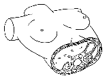
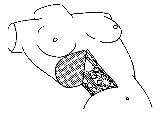
- sagittal (longitudal) - a scanning plane, when a long axis of a probe is oriented in the direction head-legs of a patient;
- frontal - a scanning plane, when a probe is located on the lateral surface of a patient's body with orientation of its long axis in the head-legs direction;
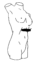
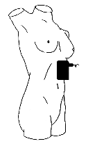
- transverse - a scanning plane, when a long axis of a probe is oriented perpendicularly to the long axis of a patient's body.
Longitudal scanning
Transversal scanning