Ultrasonic scanning.
The settings of us-scanning, determined in the software by a user, are of extreme importance if you want to get a qualitative us-image. To simplify the adjusting of many scan settings with the use of various probes and examination types there exist the SCHEMES OF SETTINGS (Fig. 9) which allow to set the parameters not separately but as a complex. Thus, for example, there are such preset schemes as CARDIO-M, ABDOMINAL EXAMINATION, GYNECOLOGY, etc. You can also add personal schemes of settings.
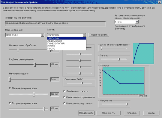
Fig. 9. Preset Scanning Schemes.
Table 2. Main technical specifications of scanning module.
| Parameter | Value |
|---|---|
| In a Scan Mode: | |
| Imaging modes | B, B+B, B+M, M. |
| Number of simultaneously connected probes | Two. Program switch |
| Max scan depth | 90-220mm. Depends on the probe type. |
| Imaging Shift from Probe Surface | Up to 40mm. Depends on probe type and scan mode. |
| Frame Average | 0% - 90%. |
| Zoom in Image Area | Fragment 125x125pix. 4 times enlargement. Colour contrast of the enlarged area. Possibility of measurements in the enlarged area. |
| Image Brightness & Contrast | Time Gain Control in 5 depth zones. · Changeable "black level". Continuously adjustable dynamic range. Gamma-correction.· Adjustable radiation power. |
| Focusing | One/two focal zones with continuously changeable focus zones. |
| Scan Density | 128 or 189 beams per frame. Program switch. |
| Image Orientation | Program adjustment LEFT/RIGHT, UP/DOWN. |
| M-Mode Specifications | Adjustable rate of M-scanning in M and B+M modes (200, 100, 50 lines/sec). Choice of M-line location. |
| High-Pass Filter | Personal preset for each mode. |
| Cineloop | Last 128 cadres record. Ability of reproduction, frame-accurate drop, record to hard disc as a reel. |
| In a Freeze Mode: | |
| Measurements | B, B+B Modes:
|
| Image Storage | Up to ten each scan session. |
| Measurement Results Report | With possibility of naming ("beparietal diameter of the fetal head", "biacromial diameter of the left kidney" etc.)and subsequent automatic inclusion into report text. |
| Automatic Calculations | Changeable obstetrics standard, calculation of major indexes, comparison to standards, inclusion of results into report text |
| Additional | Colour contrasting indicators of the details of the image, body markers with the probe distribution in the investigated part of the body |
The control of the scan modes, adjustment of the separate parameters, change for the freeze mode and back is realized with the help of a mouse or a trackball. The intelligible icons (Table 3) simplify the training and subsequent operating.
Table 3. Some of the Icons on the Control Panel.
| Icon | Function |
|---|---|
| Measurement of dimensions | |
| Choice between scan modes | |
 |
Change of brightness/contrast of the image |
The examples of the screens in different scan modes are represented in Table 4.
Table 4. General View of the Screen in Different Scan Modes
| View of the Screen | Scan Mode |
|---|---|
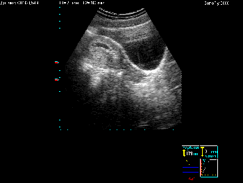 |
Mode B |
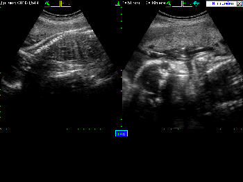 |
Mode B+B |
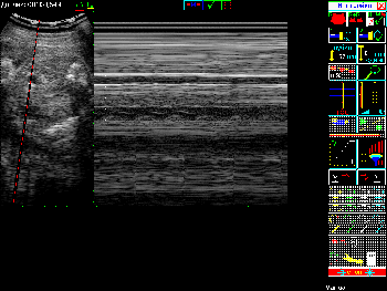 |
Mode B+M |
To indicate the name of necessary measurements with the help of simple methods is enough to realize
automatically the corresponding calculation and watch its results.
The order of the calculation results reception is as follows:
- Measurement of the parameter in the corresponding projection.
- Naming of the parameter – correspondence of a sensible phrase like "Fetus’s thigh length" or "transversal dimensions of the gallbladder" to an abstract number.
- Observation of the calculations results with the condition that you entered all necessary measurements data and they are correct.
This order is represented on Scheme 2.
1. Measurements of Internal Diameter of Ovum
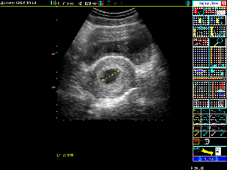
2. Record of Measurements Results
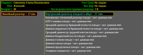
3. Observation of Calculation Results
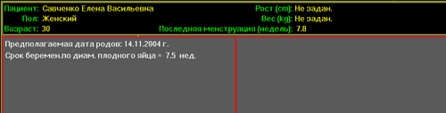
Scheme 2. Reception Order of Results Calculating Pregnancy Duration
The peculiarities of how to prepare an examination report are shown for the specialists in different branches in such sections:
Thus, the result of examination contains the created by the program and, if necessary,
added by a doctor text examination report with connected us-images. They can be stored together with
information about a patient during unlimited time period and can be analyzed and corrected any moment
you like.
SonoFly software is constantly updated, getting new possibilities. Follow our news.